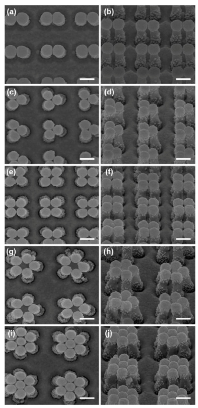
Figure 39.

SEM image showing the top view (a) and the side view with a 45° angle from the normal (b) for digons. Analogous images are reported for trigon (c,d), tetragon (e,f), pentagon (g,h), and hexagon (i,j) structures. Scale bars in the SEM images are 200 nm. Reproduced with permission from Ou et al. [358]. Copyright (2011), the American Chemical Society.
