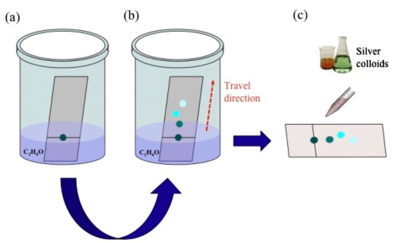
Figure 41.
(a) A drop of plasma blood containing apomorphine is put on a thin layer chromatography (TLC) slide; (b) elution with ethanol; (c) a silver colloid solution is dropped on the spots after separation has occurred. Reproduced with permission from Lucotti et al. [432]. Crown copyright (2012), published by Elsevier B.V.

