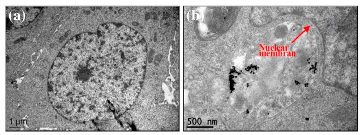
Figure 50.
(a) TEM image of human breast cancer cell, of approximately 10 µm diameter, showing cell structures, like nucleus and nuclear membrane; (b) TEM image of cell incubated with gold nanoparticles, which reside in cytoplasm and are enveloped into some vesicles (“lick up vesicles”); gold nanoparticles are clearly aggregated. Reproduced with permission from Zhu et al. [456] under Creative Commons 2.0 license (https://creativecommons.org/licenses/by/2.0/).

