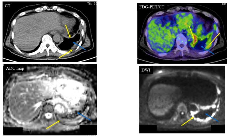
Figure 1.
Case 1—Malignant Pleural Mesothelioma (MPM). A 64-year-old male with MPM (cT4N2M0). The yellow arrows indicate the MPM. The blue arrows indicate pleural effusion. Computed tomography (CT) showed left pleural thickness of the MPM. The apparent diffusion coefficient (ADC) of the MPM was 0.84 × 10⁻³ mm2/s (positive) and the ADC of the pleural fluid was 3.95 × 10⁻³ mm2/s (negative). Fluorodeoxyglucose (FDG)-position emission tomography (PET)/CT showed partial accumulation (standardized uptake value (SUV)max: 12.39) of FDG on the MPM.

