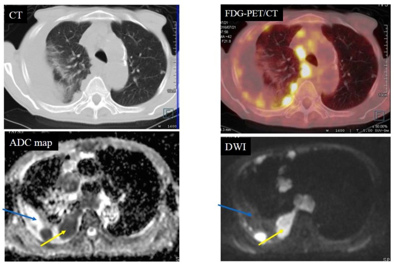
Figure 2.
Case 2—Pleural dissemination of lung cancer. An 81-year-old male with pleural dissemination of a large cell neuroendocrine carcinoma. The yellow arrows indicate pleural dissemination. The blue arrows indicate pleural fluid. The apparent diffusion coefficient (ADC) of the pleural dissemination was 0.67 × 10⁻³ mm2/s (positive) and the ADC of the pleural fluid was 3.03 × 10⁻³ mm2/s (negative). Fluorodeoxyglucose-position emission tomography/computed tomography (FDG-PET/CT) showed scattered accumulation (standardized uptake value (SUVmax): 14.7) of the FDG on the pleural dissemination.

