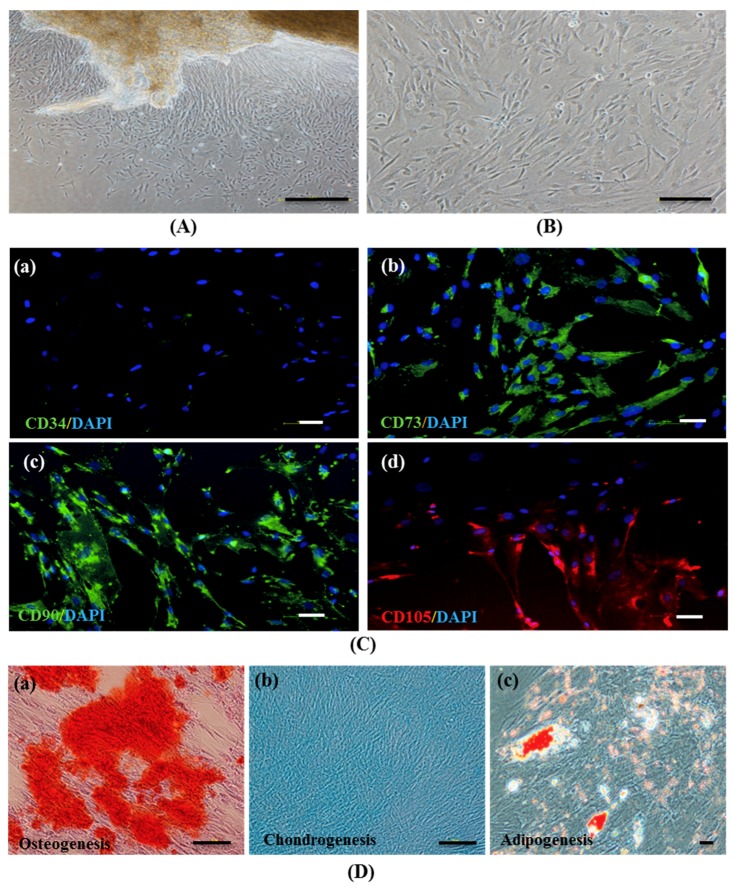
Figure 1.
The establishment and characterization of human Wharton’s jelly–derived mesenchymal stem cells (hWJ–MSCs). (A) Phase–contrast microscopic images of hWJ–MSCs were expanded from Wharton’s jelly tissue and (B) hWJ–MSCs at 80% confluence. (C) Representative images of the immunophenotype of hWJ–MSCs, as assessed for CD34 (a), CD73 (b), CD90 (c), and CD105 (d) staining. (D) Multi–lineage differentiation potential of hWJ–MSCs after 21 days, evaluated via Alizarin Red (osteogenesis) (a), Alcian Blue (chondrogenesis) (b), and Oil Red O (adipogenesis) (c) staining. (Original magnifications = 40×, bar = 200 μm (A), 100×, bar = 100 μm (B,C (a–d), and D (a,b)) and 200×, bar = 50 μm (D (c)).

