Figure 3.
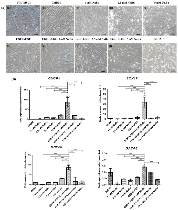
The morphological changes and real–time polymerase chain reaction (RT–PCR) analysis of mesendodermal and endodermal specific gene expressions of human Wharton’s jelly–derived mesenchymal stem cells (hWJ–MSCs) for 3 days of differentiation. (A) hWJ–MSCs were cultured with 0–5 mM sodium butyrate (NaBu) with and without epidermal growth factor (EGF) and basic fibroblast growth factor (bFGF) supplementation for 3 days. Phase–contrast microscopic images of hWJ–MSCs morphology changing after exposing 0–5 mM NaBu with and without EGF and bFGF supplementation for 3 days (b–i). hWJ–MSCs and NIH3T3 cells were used as negative and positive control cells (a,j). (Original magnifications 200×, bar = 50 μm). (B) RT–PCR analysis of definitive endoderm specific gene expression of CXCR4, SOX17, and HNF3β and mesendoderm specific gene expression of GATA6 after 3 days of pre–treatment. Gene expression was normalized to GAPDH and was shown as expression fold–change relative to that of control cells. The experiments were performed in triplicate. The data are shown as mean ± SD, * p < 0.05, and *** p < 0.001 compared to the control group. ∆, represents the significantly highest mRNA expression (p < 0.001) compared to all other groups. ## p < 0.01, and ### p < 0.001 compared to the significantly highest mRNA expression group.

