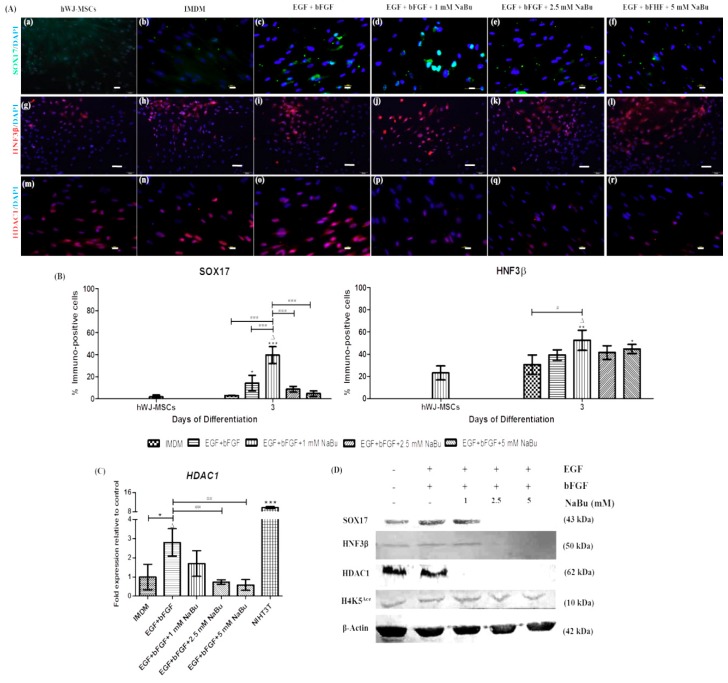
Figure 4.
The influence of sodium butyrate (NaBu) along with the epidermal growth factor (EGF) and basic fibroblast growth factor (bFGF) on the definitive endodermal differentiation capacity of human Wharton’s jelly–derived mesenchymal stem cells (hWJ–MSCs). (A) Immunofluorescence staining of the definitive endoderm and histone deacetylase type 1 (HDAC1) proteins of hWJ–MSC for 3 days of differentiation. Representative indirect immunofluorescence staining for SOX17 (a–f), HNF3β (g–l), and HDAC1 (m–r) after NaBu (0, 1, 2.5, and 5 mM) pre–treatments combined with EGF and bFGF for 3 days. (Original magnifications 400×, bar = 10 μm (a–f,m–r) and 200×, 50 μm (g–l). (B) Immunopositive cells ratio of the definitive endoderm–like cell–derived hWJ–MSCs in various NaBu induction groups and the controls for 3 days. Overall, the 1 mM NaBu condition displayed significantly higher immunopositive cells in all definitive endodermal markers than other conditions. SOX17 and HNF3β represent definitive endodermal markers. All treatments were enumerated in five different fields (n = 5). The data are shown as mean ± SD, * p < 0.05, ** p < 0.01, and *** p < 0.001 when compared to the control group. ∆, represents significantly higher protein expression (p < 0.001) than that in all other groups. # p < 0.05, and ### p < 0.001 compared to the highest protein expression group. (C) Real time–polymerase chain reaction (RT–PCR) analysis of HDAC1 gene expression after 3 days of pre–treatment. The gene expression was normalized to β–ACTIN and was calculated as an expression fold change relative to control cells. NIH3T3 cells were used as a positive control. The experiments were performed in triplicate. The data are s hown as the mean ± SD, * p < 0.05 compared to the control group. ∆, represents significantly higher mRNA expression (p < 0.01) compared to all other groups. ## p < 0.01 compared to the significantly highest mRNA expression group. (D) Western blot analysis of SOX17, HNF3β, HDAC1 and H4K5Ace protein levels after pre-treatment for 3 days; β–actin was used as an internal control.

