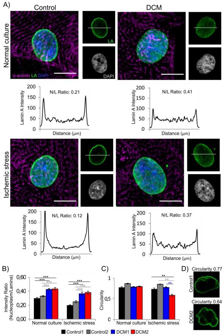
Figure 1.
Characterization of lamina structure in human induced pluripotent stem cell (hiPSC) derived cardiomyocytes. (A) Dissociated control and dilated cardiomyopathy (DCM) hiPSC-cardiomyocytes (CMs) were cultured either in normal culture conditions or exposed to ischemic stress for 3 h, fixed and stained for lamin A (LA, green), α-actinin (magenta) and DNA (DAPI; blue). Representative maximum projections of Z-stack sections (merged) and single mid-plane confocal sections (LA, DAPI) from control1 and DCM2 CMs are shown. Scale bar 10 µm. Fluorescent intensity values are illustrated below the image with nucleoplasm/lamina (N/L) ratio numbers. (B) Fluorescence intensities at the lamina region and in the nucleoplasm were determined from mid-plane confocal sections of 20–30 randomly selected cells and the average ratios of the signals (nucleoplasm/lamina) were plotted. (***) p < 0.001. (C) Quantification of nuclear circularity. (**) p < 0.01 and (***) p < 0.001. (D) Example images of different circularity values corresponding to the calculated average values (Perfect circle = 1.0).

