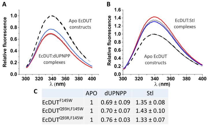
Figure 5.
The active site tryptophan sensor reports on ligand binding to EcDUT. (A) Binding of the substrate analogue dUPNPP. (B) Binding of protein Stl. (C) Comparison of peak relative fluorescence values. Black dashed lines stand for the EcDUTF145W, EcDUTQ93H,F145W and EcDUTQ93R,F145W apoenzyme constructs. Ligand bound EcDUTF145W, EcDUTQ93H,F145W and EcDUTQ93R,F145W are represented by grey, red and blue straight lines, respectively.

