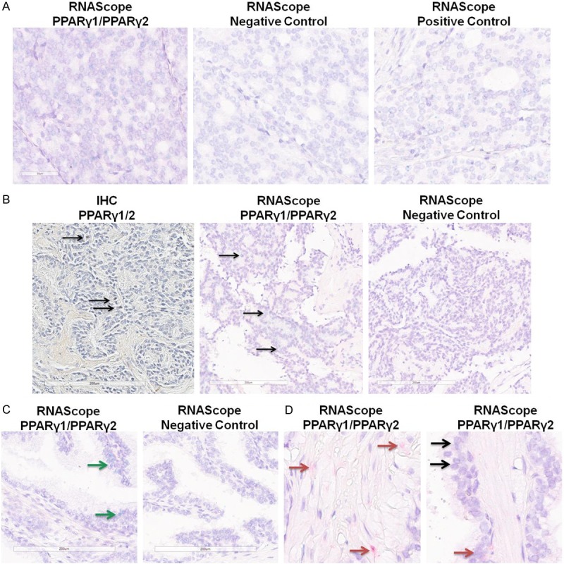
Figure 2.

Detection of PPARγ1 and γ2 RNAs in human prostate tissue. RNAs specific to PPARγ1 (green) or PPARγ2 (red) were detected using the RNAScope duplex staining platform. A. Example of staining in a PC section, demonstrating PPARγ1 staining (green) expression but no PPARγ2 staining (left), along with negative and positive control staining on adjacent sections. B. Example of staining in a metastatic PC sample. (Left) IHC staining with the antibody that detects both PPARγ1/2 isoforms. (Center) RNAScope staining for PPARγ1 and PPARγ showing only PPARγ1 staining. (Right) RNAScope negative control staining. Arrows show positive nuclei. C. Example of RNAScope staining for PPARγ1 and PPARγ2 showing only PPARγ1 staining in a benign gland (left) with negative control (right). Green arrows show PPARγ1 staining. D. Examples of RNAScope staining for PPARγ1 and PPARγ2 showing PPARγ2 staining in stroma (left) and PPARγ1 and PPARγ2 in benign epithelial cells (right). Red arrows show PPARγ2, black arrows show areas of both PPARγ1 staining (green) and PPARγ2 staining (red).
