Figure 2.
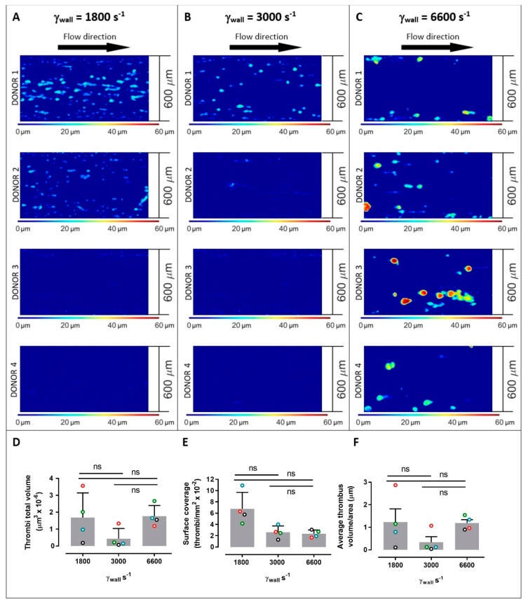
Platelet thrombus formation in straight channels. Images of thrombus formation (demonstrated by contours of thrombus height) all collected under the same conditions from anticoagulated blood from 4 separate donors within collagen-coated channels with constant wall shear rates (γw) of 1800 s−1 (A), 3000 s−1 (B), and 6600 s−1 (C) (donors 1–4 are shown from top to bottom panels at each shear rate). Measured thrombi total volume (D), surface coverage (E), and average thrombus volume/area (F) for 4 donors (open circles; red = donor 1, green = donor 2, blue = donor 3, black = donor 4) at 1800 s−1, 3000 s−1, and 6600 s−1, where bars represent mean ± standard deviation. Data in panels (D–F) were evaluated using one-way ANOVA with Tukey’s correction for multiple comparisons. ns = no significance. Larger numbers of smaller thrombi formed at 1800 s−1, while there were lower numbers of larger thrombi at 6600 s−1, and at the intermediate wall shear rate of 3000 s−1, thrombus formation was generally reduced suggesting distinct shear-dependent mechanisms regulating initial adhesion or growth of thrombi. Despite the differences in thrombus geometries, the total volumes of immobilised platelets at 1800 and 6600 s−1 were comparable (A).

