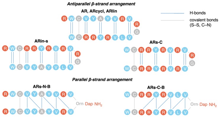
Figure 3.
Different hydrogen bonding patterns observed for arenicin variants. AR, ARin-s, and ARs-C show the same H-bonding pattern (dotted blue lines) typical of antiparallel β-strands (two H-bonds connect the α-amine and α-carboxyl groups of an amino acid on one side of the hairpin with the α-amine and α-carboxyl groups of an amino acid on the other side of the hairpin). ARs-N-B and ARs-C-B instead show the pattern typical of parallel β-strands (two H-bonds connect the α-amine and α-carboxyl groups of an amino acid on one side of the hairpin with the α-amine and α-carboxyl groups of two different amino acids on the other side). The disulfide bond is indicated by a solid grey line, as well as branching from the ornithine residue α- and δ-amines (Figure 7).

