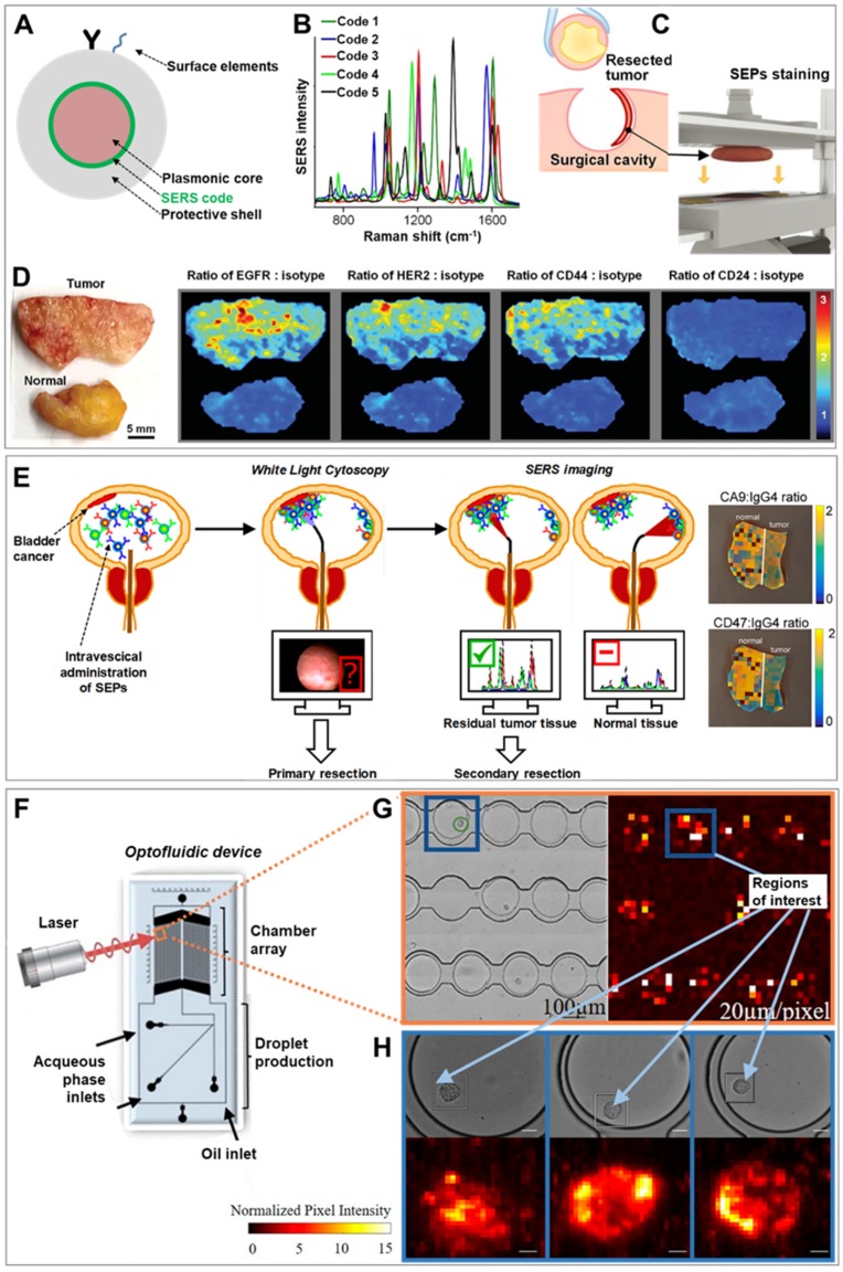
Figure 1.
(A) Schematic depiction of the traditional surface-enhanced Raman scattering (SERS)-encoded nanoparticle (SEP) construction. (B–D) Application of SERS imaging to resected tumor tissue for intraoperative surgical guidance: (B) Unique SERS spectra of five different codes on gold particles coated with a silica shell. (C) Upon excision, the resected sample is placed in an automated staining device combining multiple quick dipping of the specimen into a SEPs solution with high-frequency vibration for fast and extensive topically applications of encoded particles onto the surface of the fresh tissue. (D) Ratiometric images biomarker targeting SEPs vs. negative control (isotype) obtained from raster-scanned SERS imaging (<3 min) of the illustrated human breast tumor and normal tissue. Adapted with permission from [18]. Copyright 2016, Wiley-VHC. (E) Schematic of the proposed application of intravesical SERS imaging for intraoperative endoscopic surgery. Adapted with permission from [19]. Copyright 2018, American Chemical Society. (F–H) SERS-microfluidic device for single live cell analysis: (F) Schematic depiction of the droplet-based optofluidic device; (G) Low-resolution map of the chamber array, (H) High-resolution map of individual cells encapsulated in droplets. Adapted with permission from [20] Copyright 2018, American Chemical Society.

