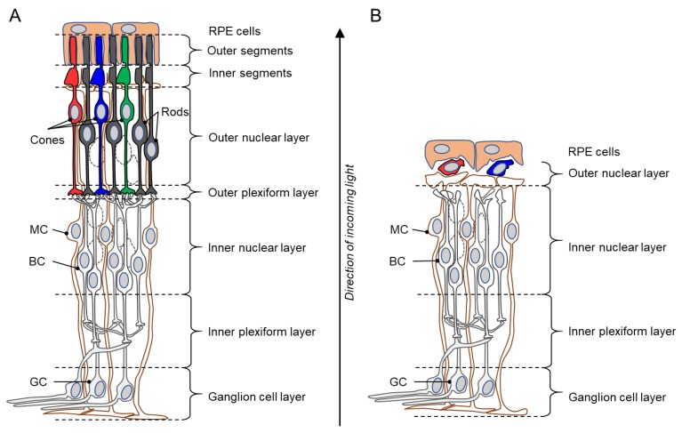
Figure 1.
Schematic drawings of a healthy retina and a inherited retinal degeneration (IRD) retina. (A) Illustration of the various layers of an intact, healthy retina, from the retinal pigment epithelium (RPE) to the ganglion cell layer. Rod photoreceptors in the outer retina are shown in black, while cones are indicated by red, green, and blue. (B) Degenerated IRD retina. The outer nuclear layer is almost completely lost and the outer plexiform layer has essentially disappeared. Curiously, when the retina has lost all functionality, a small number of cone photoreceptors may still be present, possibly for many years beyond the loss of rod photoreceptors. BC = bipolar cell; GC = ganglion cell; MC = Müller glial cell. Note that the retinal structure has been simplified for clarity and that not all retinal cell types are shown.

