Figure 4.
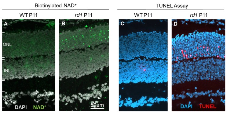
Incorporation of NAD+ as a biomarker for retinal cell death. At post-natal day 11 (P11), when compared to wild-type (WT) retina (A), photoreceptors in the rd1 mouse model (B) display marked incorporation of biotinylated NAD+. This is highly correlated with the TUNEL assay for cell death, which at the same age detects only few cells in the WT situation (C), while large numbers are detected in the rd1 outer nuclear layer (ONL; D). INL = inner nuclear layer, GCL = ganglion cell layer.

