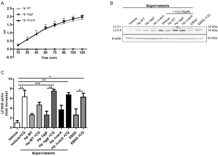
Figure 2.
Hp∆ggt culture supernatant increases autophagic flux of AGS cells in comparison with the parental strain. (A) Activity of gamma-glutamyltranspeptidase in cell lysates of wild type (HpWT) and the isogenic Hp∆ggt and Hp∆vacA mutants (lacking gamma-glutamyltranspeptidase and vacuolating toxin, respectively). (B) AGS cells were incubated for 6 h with the concentrated supernatants from wild type, Hp∆ggt and Hp∆vacA H. pylori strains (1:25) in the presence or absence of chloroquine (30 μM). As a control, concentrated non-conditioned medium (vehicle) was added at a similar dilution. In addition, Earle’s Balanced Salt Solution medium (amino acid and serum free medium) was used as a positive control to induce autophagy (4 h). Protein levels of the microtubule-associated protein 1A/1B light chain 3 (LC3) conjugated to phosphatidylethanolamine (LC3-II) and β-actin were evaluated by Western blotting. In panel C) the quantification of relative levels of LC3-II for AGS cells is shown. These data represent the mean ± the standard error of the mean (SEM) of three independent experiments. * p < 0.05, ** p < 0.01, *** p < 0.001.

