Figure 3.
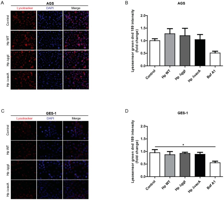
H. pylori infection did not affect lysosomal acidification in gastric cells. AGS and GES-1 cells were infected with H. pylori wild type (HpWT) or the isogenic Hp∆ggt and Hp∆vacA (lacking gamma-glutamyltranspeptidase and vacuolating toxin, respectively) mutant strains at a multiplicity of infection (MOI) of 100 for 6 h. (A) To visualize acidified compartments, (A) AGS and (C) GES-1 cells were treated with the acidotropic dye Lysotracker Red DND-99 (75 nM) for 2 h. Representative fluorescent images from three independent infection experiments are shown. To measure the lysosomal pH, (B) AGS and (D) GES-1 cells were treated with Lysosensor green DND-189 (1 µM) for 30 min. Fluorescence intensity was evaluated by flow cytometry. Bafilomycin A1 (100 nM) was used as positive control to inhibit acidification. Data represent the mean ± the standard error of the mean (SEM) of at least three independent experiments. * p < 0.05.

