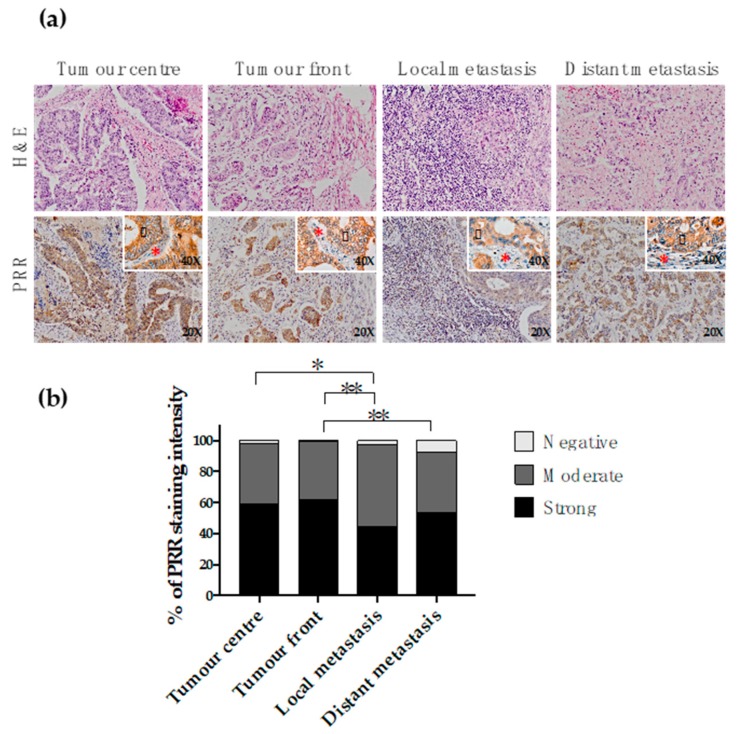
Figure 2.
Immunohistochemical PRR staining along the conversion of the primary tumour into metastasis. (a) Representative fractions of the centre of the primary tumour (n = 228), infiltrating front of the primary tumour (n = 222), local lymph node metastasis (n = 176) and distant metastasis (n = 94) were immunohistochemically stained with an antibody against PRR. Granular cytoplasmic staining was observed in neoplastic cells (black arrowhead). Stromal cellularity (lymphocytes and fibroblasts) did not express PRR (red asterisk). (b) PRR staining intensity was scored as negative, moderate or strong. The scores were quantified in each tissue type and statistical significance of the PRR intensity pattern among the different tissues was determined by Chi-Square test. H&E: Hematoxylin and Eosin staining. PRR: (pro)renin receptor staining.

