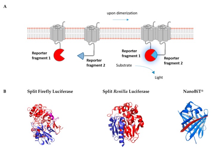
Figure 4.
Luminescence-based protein complementation assay. (A) The principle of the luminescent complementation assay is shown schematically. (B) Split versions of the luminescent reporters Firefly, Renilla, and Nanoluciferase are shown in blue and red. Purple refers to overlapping sections. Split luminescent biosensors are depicted in proportion to their size (Fluc: 62 kDa, Rluc: 36 kDa, and NanoBiT®: 19 kDa) (PDB Accession no. 1LCI for Firefly luciferase, PDB Accession no. 2PSD for Renilla luciferase) (NanoBiT®: source Promega).

