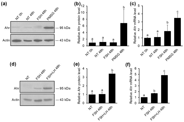
Figure 1.
The effect of gonadotropins on Ahr expression in ovarian GCs in vivo. (a) Mice were injected once with 5 IU of PMSG, 5 IU of FSH or vehicle (NT) and lysates were collected from granulosa cells (GCs) isolated before (0 h) or 48 h later. Representative Western blot shows Ahr and actin protein levels. Specificity of Ahr antibody is shown in Supplementary Figure S1d). (b) Densitometry analysis of three independent experiments (mean ± SD) representing Ahr protein levels normalized to actin. (c) qPCR analysis of Ahr mRNA levels. (d) Mice were injected in total 4 times (every 12 h) with FSH (1.5 IU) or FSH (1.5 IU) + LH (1.25 IU) and lysates were collected from GCs isolated before (NT) or 48 h after initial injection. Representative Western blot shows Ahr and actin protein levels. (e) Densitometry analysis of three independent experiments (mean ± SD) representing Ahr protein levels normalized to actin. (f) qPCR analysis of Ahr mRNA levels. Bars with no common superscripts are significantly different (p < 0.05).

