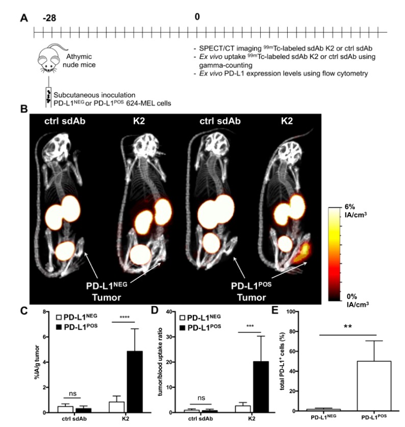
Figure 4.
Radiolabeled sdAb K2 allows visualization of human PD-L1POS melanoma tumors by nuclear imaging. (A) Scheme of the experimental setup. (B) SPECT/CT images showing the biodistribution of 99mTc-K2 or 99mTc-R3B23 (control sdAb) one hour after i.v. administration in athymic nude mice bearing PD-L1NEG (left) or PD-L1POS (right) 624-MEL tumors (n = 6). (C,D) Ex vivo analysis of the accumulation of 99mTc-sdAbs in dissected PD-L1NEG or PD-L1POS 624-MEL tumors (C, expressed as %IA/g), and of tumor-to-blood uptake ratios (D), 80 min after i.v. radiotracer injection (n = 6). (E) Percentage of human PD-L1POS cells in tumors dissected from mice that were s.c. implanted with parental 624-MEL cells (PD-L1NEG) or PD-L1-modified counterparts (PD-L1POS), as measured by flow cytometry analysis of tumor single cell suspensions (n = 6). ** p < 0.01, *** p < 0.001, **** p < 0.0001.

