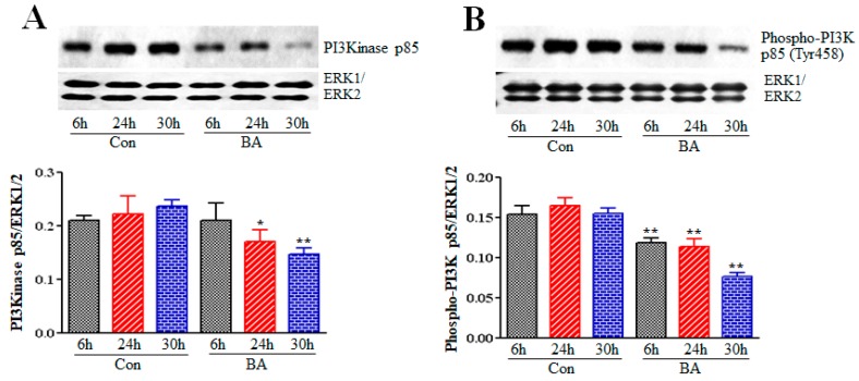
Figure 2.
Effect of butyrate (BA) on PI3K in VSMCs. Proliferating VSMCs were treated with or without 5 mM butyrate for indicated periods of time. At the end of treatment, cell lysates were prepared and subjected to Western blot analysis to determine the PI3K expression and activation level by measuring the level of unphosphorylated and phosphorylated p85 subunit of PI3K, respectively. Immunoblotting of ERK1/2 was performed with the same lysate to normalize the protein loading. The band intensities were measured and normalized to protein loading. The data obtained were analyzed and presented as mean ± S.D. (A) Expression level of PI3K p85 subunit measured by using antibody specific to p85 subunit (* p < 0.01 vs 24 h control (Con), and ** p < 0.001 vs 30 h control (Con). (B) Activation of p85 subunit of PI3K was evaluated by the antibody specific to Tyr458-phosphorylated p85 (** p< 0.001 vs respective control (Con).

