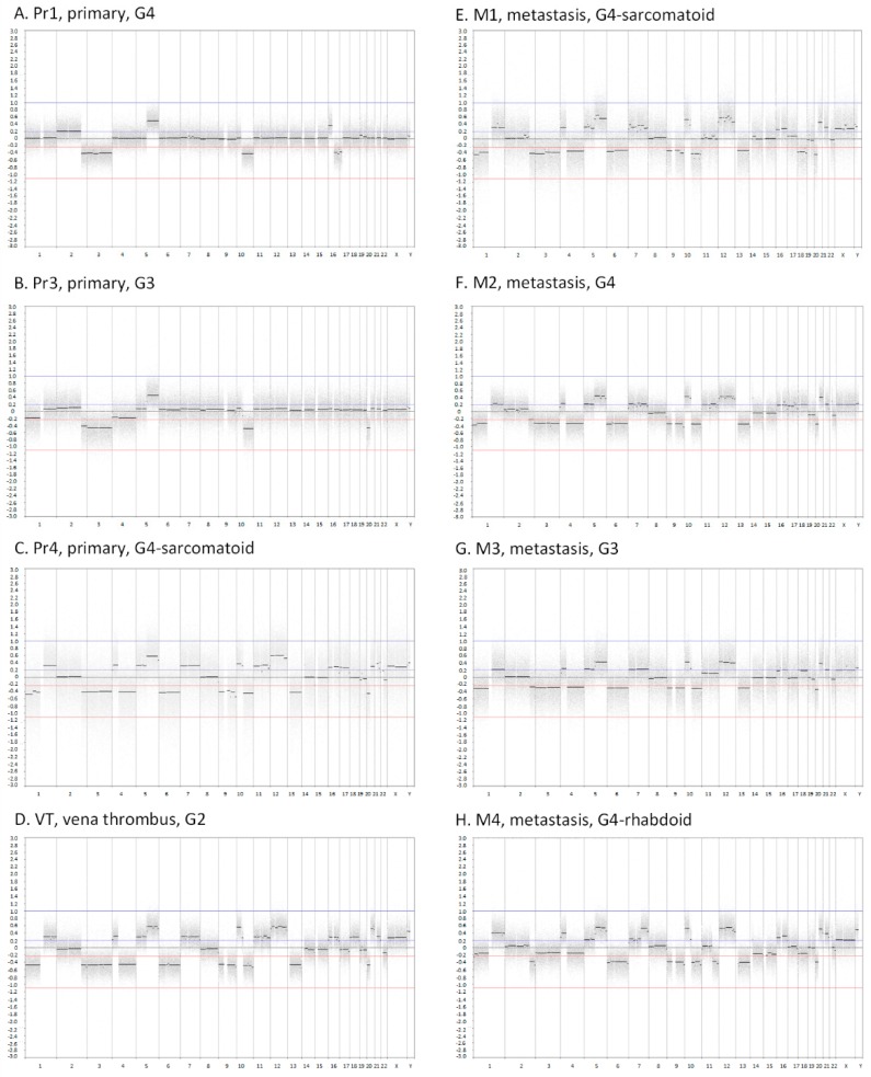
Figure 1.
Array comparative genomic hybridization (CGH) plots of eight tumour samples from ccRCC patient 1 (RC1). The x-axes show the genomic position starting from 1pter until Xqter. The y-axes indicate the log2 intensity ratio between tumour and reference. Abbreviations: Pr1, primary tumour 1; Pr3, primary tumour 3; Pr4, primary tumour 4; VT, inferior vena cava tumour thrombus; M1, metastasis 1; M2, metastasis 2; M3, metastasis 3; M4, metastasis 4; G1, tumour grade 1; G2, tumour grade 2; G3, tumour grade 3; G4, tumour grade 4.

