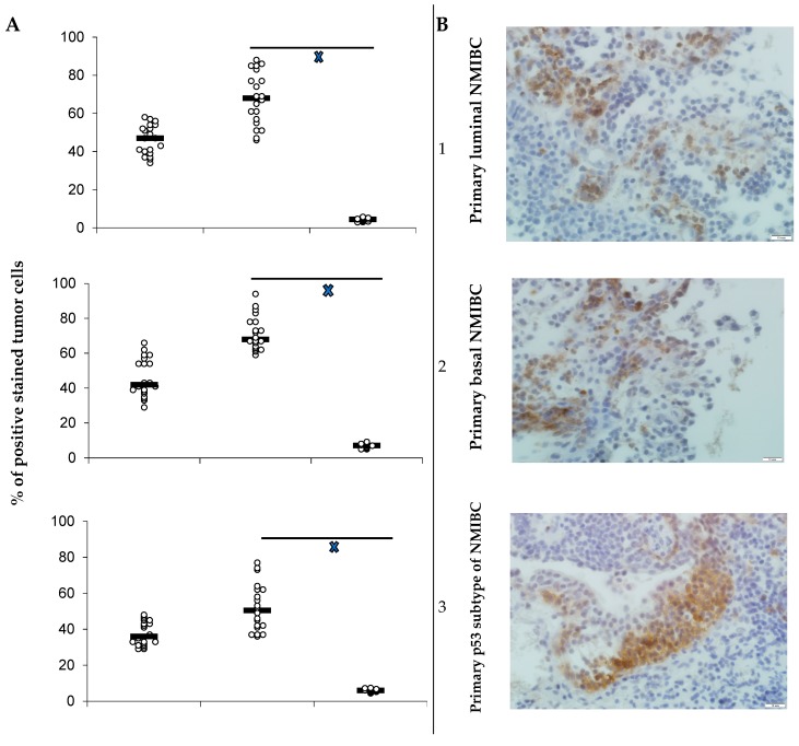
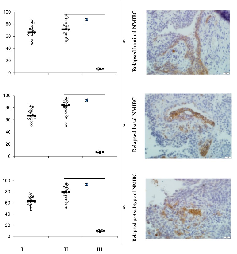
Figure 1.
(A) PD-L1 expression in bladder cancer PDXs in dependence on PD-L1 treatment; PD-L1 expression as scatterplots of individual % of positively stained tumor cells with estimated median for maternal tumor used for engraftment (I), for PDX of control subgroup mice (II), and animals (III) utilized specific therapy (n = 20 in maternal tumor group; n = 10 in each subgroup); xp < 0.05 when compared with control (Student’s t-test). (B) PD-L1-positive staining of primary luminal (1), basal (2), and p53 subtypes (3) of NMIBC, and relapsed luminal (4), basal (5), and p53 (6) subtypes of bladder cancer in maternal tumor’s specimens; IHC staining, x600.


