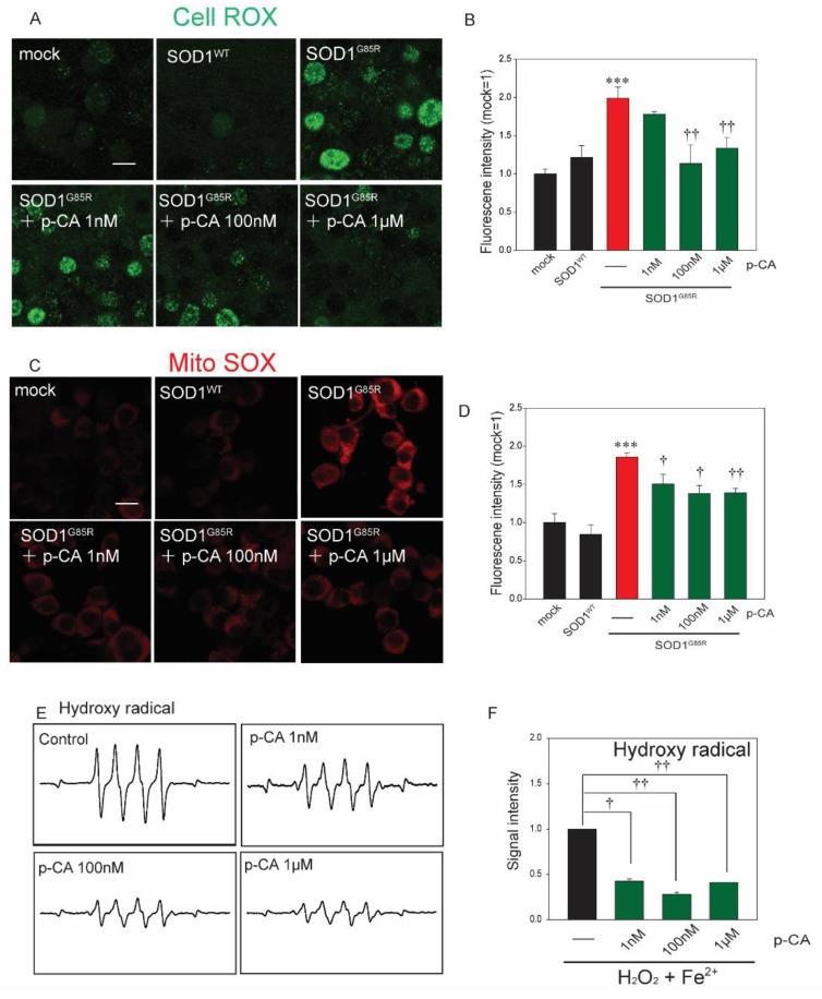
Figure 2.
p-CA reduced mutant-SOD1-related oxidative stress. (A) Confocal imaging of CellROX in N2a cells transfected with mCherry–SOD1G85R with p-CA for 24 h. (B) Quantified analysis of CellROX using Image J. (C) Confocal imaging of MitoSOX in N2a cells transfected with mCherry–SOD1G85R with p-CA for 24 h. (D) Quantified analysis of MitoSOX using Image J. (E) Typical spectra of DMPO-OH spin generated from H2O2 plus Fe2+ in the absence (control) or presence of p-CA. (F) The amount of hydroxy radicals was semi-quantitatively measured as the formation of DMPO-OH spin adducts by ESR spectrometry. Differences were evaluated by one-way ANOVA (mean ± SEM, n = 3). *** p < 0.001 vs. SOD1WT, †† p < 0.01, † p < 0.05 vs. SOD1G85R. Scale bar: 10 µm. p-CA: p-coumaric acid.

