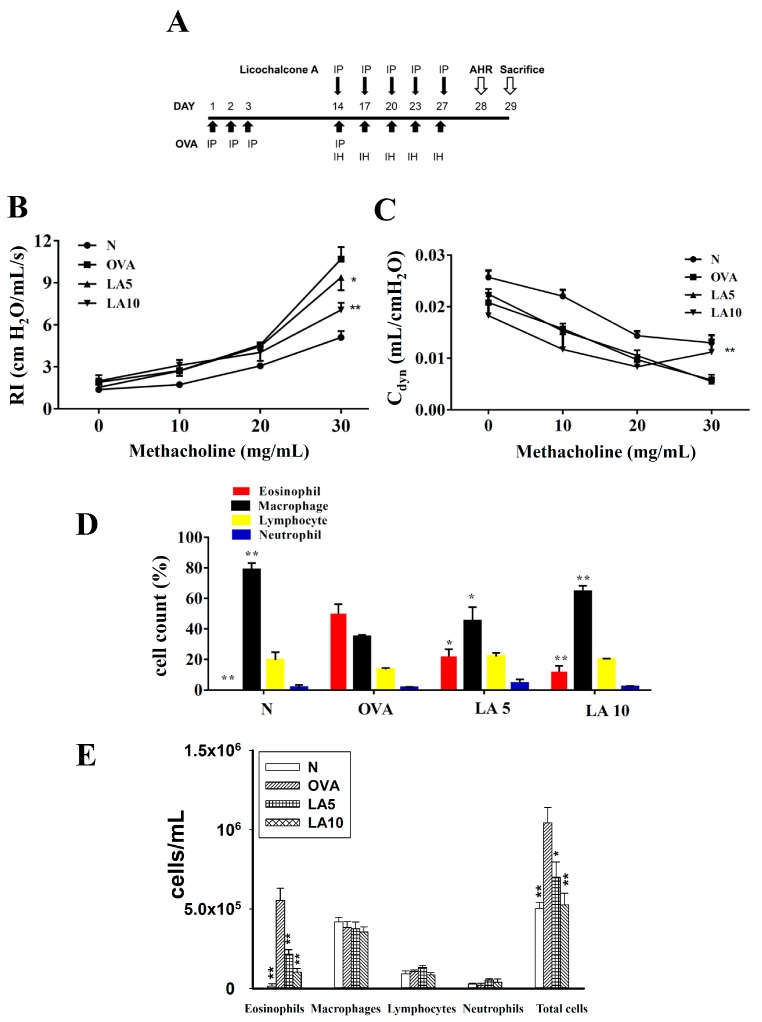
Figure 1.
The effect of licochalcone A (LA) on airway hyper-responsiveness (AHR) and cell counts in bronchoalveolar lavage fluid (BALF) of asthmatic mice. (A) On days 1–3 and 14, mice were sensitized with ovalbumin (OVA) by intraperitoneal injection (IP) and challenged with 2% OVA inhalation (IH) on days 14, 17, 20, 23, and 27. One hour before the OVA challenge or methacholine inhalation, mice were treated with LA or DMSO (n = 12 mice/group). (B) AHR was measured as a percentage of lung resistance (RI) from baseline normal (N) and (C) dynamic lung compliance (Cdyn). (D) Inflammatory cells were measured and the percentage of inflammatory cells in the BALF presented. (E) Inflammatory cells and total cells were measured in BALF. Three independent experiments were analyzed, and data were presented as mean ± SEM. * P < 0.05 compared to the OVA control group. ** P < 0.01 compared to the OVA control group.

