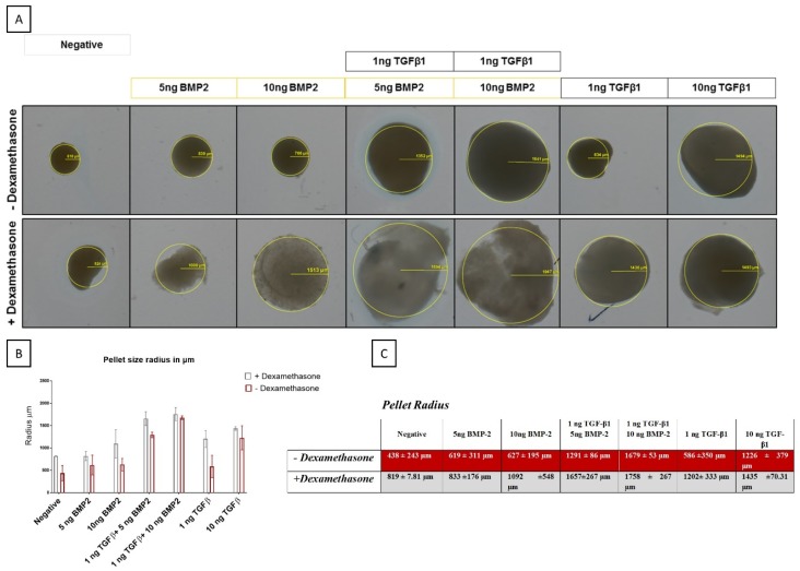
Figure 5.
Macroscopic evaluation of pellet size from hSDSCs exposed to TGF-β1 and BMP-2 in the presence or absence of dexamethasone. hSDSCs in 3D culture were cultures for 21 days in the presence of 10 ng/mL TGF-β1 (positive control) or in its absence (negative), low TGF-β1 concentration (1 ng/mL TGF-β1), various concentrations of BMP-2 alone (5 ng/mL BMP-2, 10/mL ng BMP-2) or BMP-2 in combination with 1 ng TGF-β1 (1 ng/mL TGF-β1 + 5 ng/mL BMP-2 and 1 ng/mL TGF-β1 + 10 ng/mL BMP-2). The figures are representative of four separate experiments in four different donors (A). Radius sizes are expressed in µm (B–C). Radius pellet measured with Axio Plan Microscope objective 2.5×.

