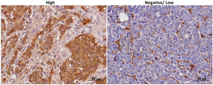
Figure 1.
Anexeleto (AXL) staining in thyroid cancer samples. Representative images of high and low/negative AXL immunohistochemical staining in thyroid cancer samples at 40× magnification, scale bar = 50 μm. High and negative/low staining was defined as described in the Section “Materials and Methods”.

