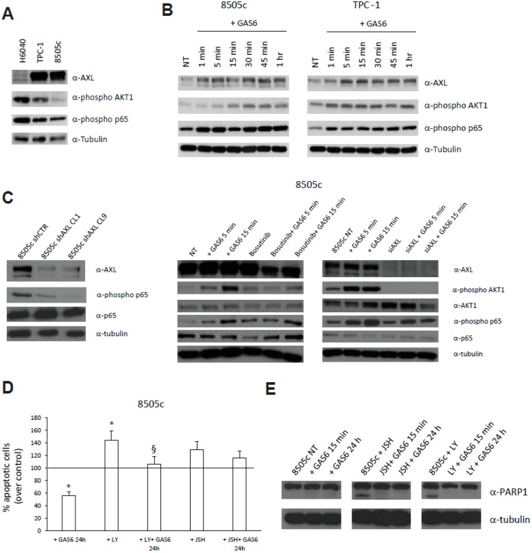
Figure 6.
In vitro evaluation of AXL functions. (A) AXL, phospho-AKT1 and phospho-p65 levels evaluated in the indicated thyroid cell lines by Western Blot (WB) analysis. Anti-tubulin antibodies served as control for equal loading. (B) AXL, phospho-AKT1 and phospho-p65 levels in 8505c and TPC-1 cells treated with GAS6 (100 ng/mL) for the indicated time points, evaluated by WB analysis. (C) Levels of AXL, phospho-p65, and p65 in 8505c cell clones stably silenced for AXL (shAXL). Two clones are shown. AXL, phospho-AKT1, AKT1, phospho-p65, and p65 levels in 8505c cells treated or not with GAS6 (100 ng/mL) for the indicated time points in the presence or absence of Bosutinib (25 μM) or siRNAs targeting AXL (siAXL). Anti-tubulin antibodies served as control for equal loading. (D) Percent relative to control of 8505c apoptotic cells, assessed by TUNEL assay, treated or not with GAS6 (100 ng/mL) in the presence or absence of LY294002 (15 μM—AKT1 inhibitor) and JSH23 (5 μM—p65 inhibitor). * p < 0.05 vs. not treated cells. §, p < 0.05 vs. the relative control. (E) PARP1 cleaved levels in 8505c cells treated or not with GAS6 (100 ng/mL) in the presence or absence of LY294002 (15 μM) and JSH23 (5 μM). Anti-tubulin antibodies served as control for equal loading.

