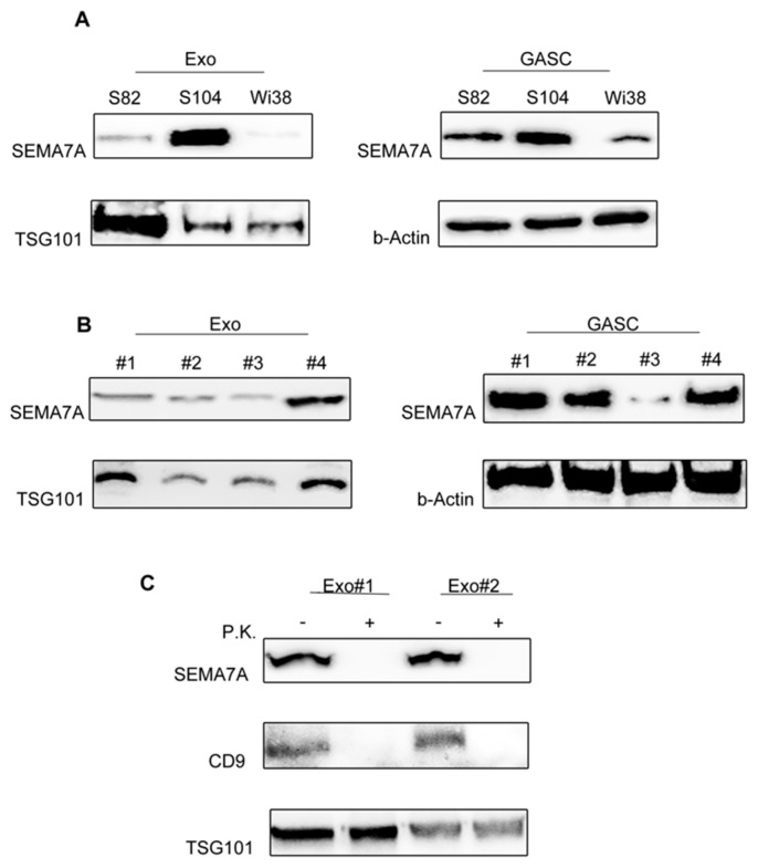
Figure 2.
SEMA7A (Semaphorin7A) is present on the surface of Glioma Associated Stem Cells (GASC)-derived exosomes. (A) Western blotting showing the expression of SEMA7A in the same exosomal preparations used for the proteomic study (Exo_S82 and Exo_S104, left panel) and in the matched producing cells (GASC_S82 and GASC_S104, right panel). As healthy control, SEMA7A expression was evaluated both in Wi38 cells and in Wi38-derived exosomes (Exo-Wi38). (B) SEMA7A expression was assessed in 4 different GASC cell lines (GASC#1, GASC#2, GASC#3 and GASC#4) and in the respective derived exosome preparations (Exo#1, Exo#2, Exo#3 and Exo#4). Membranes were hybridised with anti-TSG101 (Tumour Susceptibility Gene 1) and beta-Actin to show the reliability of exosomes and cell lysates respectively. (C) Western blot analysis for SEMA7A, CD9 and TSG101 in Exo#1 and Exo#2 preparations either untreated (−) or subjected to proteinase K treatment (P.K., +).

