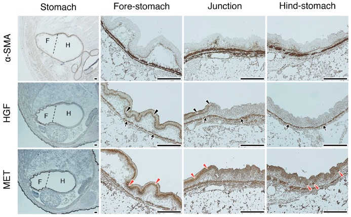
Figure 3.
Localizations of HGF and MET receptors in the developing stomach. Immunohistochemical staining was performed using t5A11 anti-human HGF monoclonal antibody or anti-MET antibody. Stomachs in day 16.5 embryos were divided into the anterior/fore-stomach and posterior/hind-stomach, distinguished by dotted lines. Black arrows indicate HGF localization in smooth muscle cells. Black arrowheads indicate HGF localized in the epithelial cells. Red arrowheads indicate MET expression in epithelial cells. Similar localization patterns were obtained in sections from two different mice. Tissues were obtained from day 16.5 embryos of hHGF-ki mice. Scale bars represent 200 µm.

