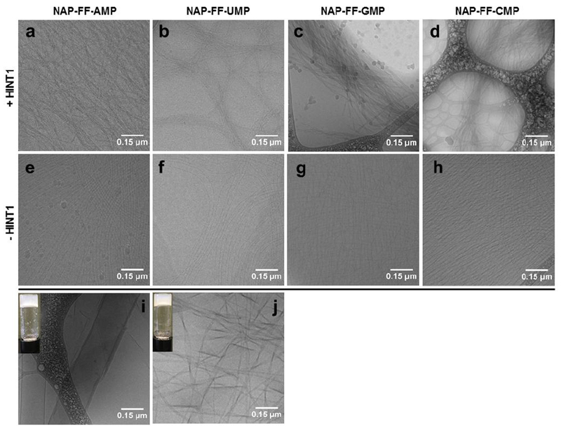
Figure 4.

Cryo-TEM of Hydrogels and Substrates. Top Panel: Hydrogels formed in the presence of HINT1 (a) NAP-FF-AMP, (b) NAP-FF-UMP, (c) NAP-FF-GMP, (d) NAP-FF-CMP. Middle Panel: Phosphoramidate Pro-Gelator Nanofibers in Buffer (e) NAP-FF-AMP, (f) NAP-FF-UMP, (g) NAP-FF-GMP, (h) NAP-FF-CMP. Bottom Panel: (i) NAP-FF-NH2 structure in the presence of PBS only, (j) and in the presence of AMP. (All scale bars represent 0.15 μM)
