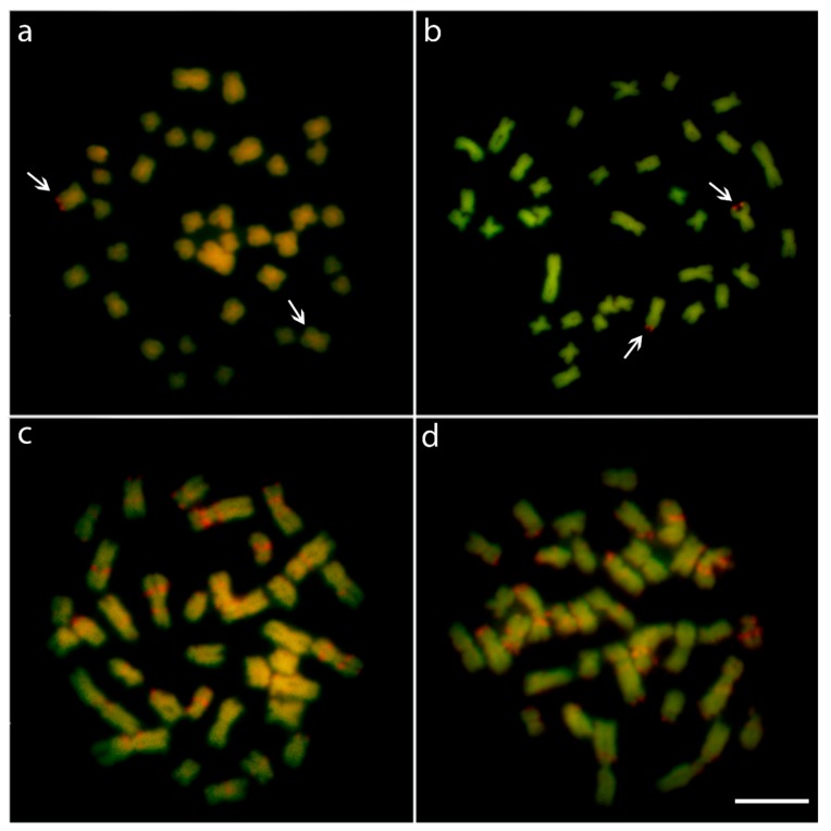
Figure 2.
Metaphase plates of male (a) and female (b) Lebiasina bimaculata and male (c) and female (d) Lebiasina melanoguttata after DAPI-CMA3 staining. The arrows indicate the unique CMA3+ site and its polymorphic state between male and females of L. bimaculata. In L. melanoguttata, males and females display a set of CMA3+ (GC-rich) and DAPI+ (AT-rich) regions on the chromosomes. Scale bar = 5 µm.

