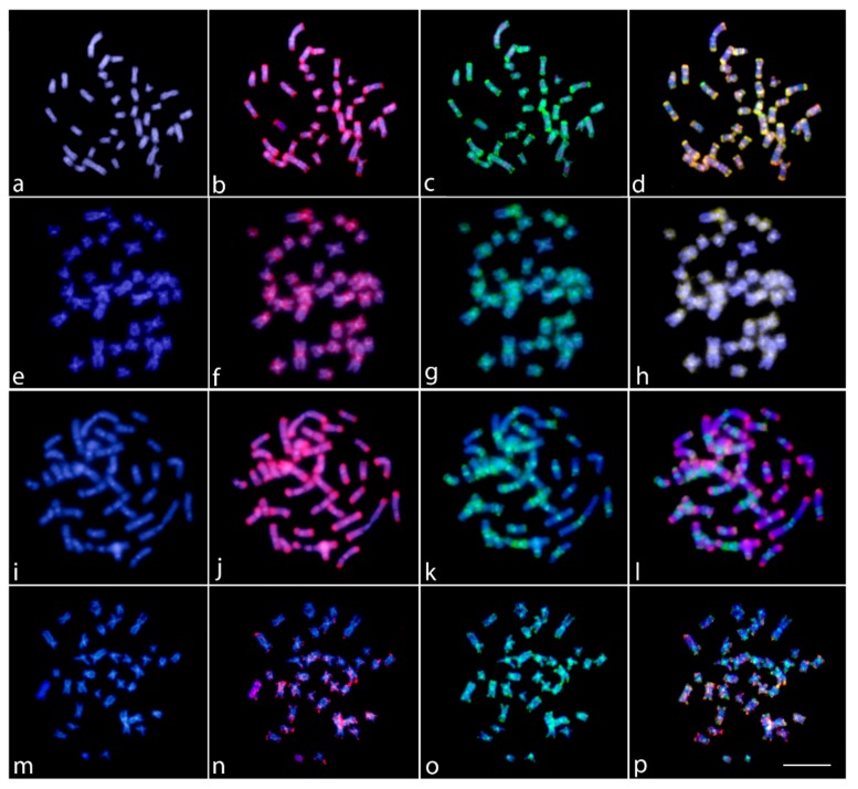
Figure 5.
Comparative genomic hybridization (CGH) for intra- and interspecific comparison in the female metaphase plates of Lebiasina bimaculata (a–d and m–p) and L. melanoguttata (e–h and i–l). Male- and female-derived genomic probes from L. bimaculata mapped against female chromosomes of L. bimaculata (a–d); Male- and female-derived genomic probes from L. melanoguttata mapped against female chromosomes of L. melanoguttata (e–h); female-derived genomic probes from both L. bimaculata and L. melanoguttata hybridized together against female chromosomes of L. melanoguttata (i–l); and female-derived genomic probes from both L. bimaculata and Boulengerella lateristriga (Ctenolucidae) hybridized together against female chromosomes of L. bimaculata (m–p). First column (a,e,i,m): DAPI images (blue); second column (b,f,j,n): hybridization patterns using male gDNA of L. bimaculata (b), male gDNA of L. melanoguttata (f), female gDNA of L. melanoguttata (j), and female gDNA of B. lateristriga probes (red); third column (c,g,k,o): hybridization patterns using female gDNA of L. bimaculata (c,o) and female gDNA of L. melanoguttata (g,k) probes (green); fourth column (d,h,l,p): merged images of both genomic probes and DAPI staining. The common genomic regions are depicted in yellow. Scale bar = 5 µm.

