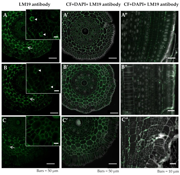
Figure 4.
Distribution of LM19 epitope in cross sections in differentiated zone (A,A’–C,C’) and longitudinal sections in meristematic zone (A”–C”) of root tissues of barley cv. Sebastian. (A–A”)—control pH 6; (B–B”)—control pH 4; (C–C”)—30 µM AlCl3. (A–C) inset—higher magnification of cortex with intercellular spaces. Detection of LM19 epitope (A–C), Calcofluor (CF), and DAPI staining with immunolocalization of LM19 epitope (A’–C’,A”–C”) are shown. (A–C)—arrowheads within the insets show fibers with LM19 epitope. (A–C)—arrows show developing intercellular spaces with LM19 epitope recognized. Epitope distribution in representative sections are shown.

