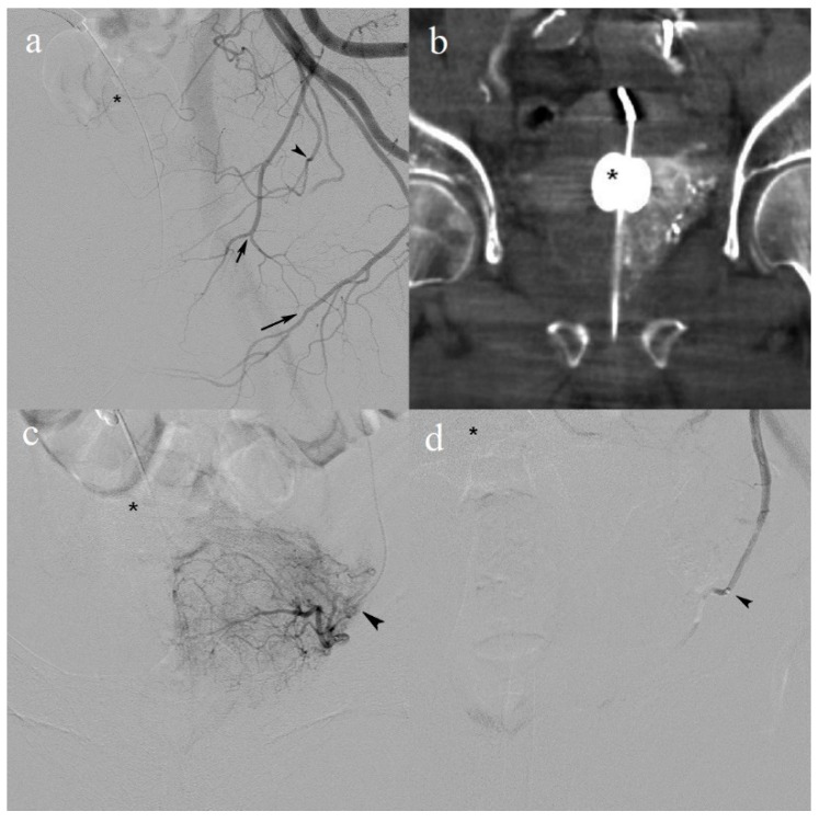
Figure 1.
(a) Selective digital subtraction angiography (DSA) in the left anterior oblique position with the catheter tip in the left internal iliac artery (not visible). The internal pudendal artery (long arrow) and the characteristic division of obturator artery (short arrow). The prostate artery (arrowhead) protrudes beneath the Foley balloon (*) catheter; (b) coronal cone-beam computed tomography with contrast enhancement in the left lobe confirming correct catheter placement; (c) frontal DSA before embolization with injection on the microcatheter in the prostate artery (arrowhead); (d) frontal DSA with the microcatheter in the same position (arrowhead) and the angiographic endpoint after embolization.

