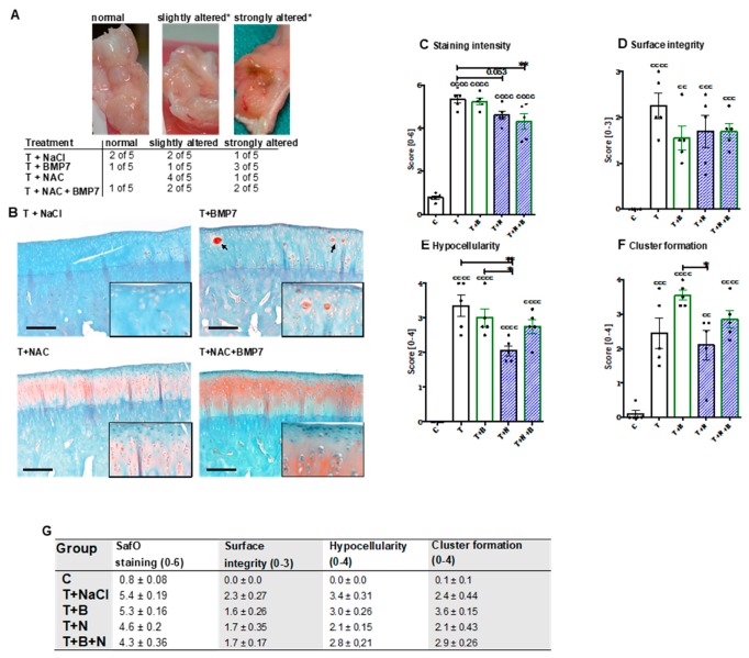
Figure 1.
Macroscopic and histopathologic assessment of the joint capsule and SafO-stained condyle sections, respectively. (A)*Alteration was defined as reddish/russet color, which was considered as possible indication of inflammatory processes or intra-articular bleeding (blood residues), and vascularization. (B) Exemplary images of SafO-stained medial condyles of each group. Cell clusters are indicated by black arrows. The black bar represents 100 µm. (C–F) Corresponding statistical analysis of the single parameters of the histopathological assessment, charted as scattered plots with bars: (C) proteoglycan staining intensity, (D) surface integrity, (E) hypocellularity, and (F) cluster formation. (G) Values of the single criteria given as mean ± SEM; n = 5. Statistically significant differences between groups were depicted as: [vs C] cc: p < 0.01, ccc: p < 0.001, and cccc: p < 0.0001; [vs. T] *: p < 0.05, and **: p < 0.01. T = trauma, T+B = traumatized and BMP7-treated, T+N = traumatized and NAC-treated, T+N+B = traumatized and BMP7- plus NAC-treated.

