Figure 4.
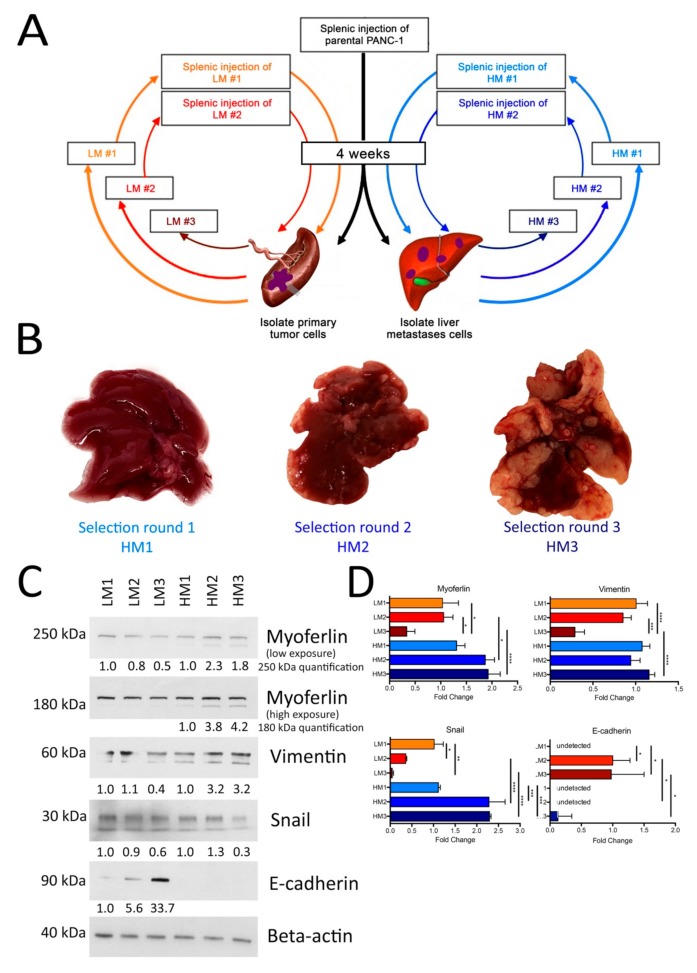
Myoferlin is overexpressed in cells with a high metastatic potential. (A) Schematic representation of the in vivo selection of liver-tropic PDAC cells used to generate low metastatic (LM) and highly metastatic (HM) clones. Adapted from [34]. (B) Representative liver from mice injected with the liver-tropic Panc-1 cells selected after one, two, or three rounds of injection. (C) Western-blot evaluation of myoferlin, vimentin, snail, and E-cadherin abundance in Panc-1 clones selected for their low (LM) or high (HM) metastatic potential. Beta-actin was used as a loading control. (D) Gene expression (mRNA) level for myoferlin, vimentin, snail, and E-cadherin in Panc-1 clones selected for their low (LM) or high (HM) metastatic potential. LM1 (LM2 for E-cadherin) gene expression was fixed to 1 and other samples were compared to LM1 (LM2 for E-cadherin). One representative experiment out of three is illustrated. Each data point represents mean ± SD, n = 3. **** P < 0.0001, *** P < 0.001, ** P < 0.01, * P < 0.05.

