Figure 4.
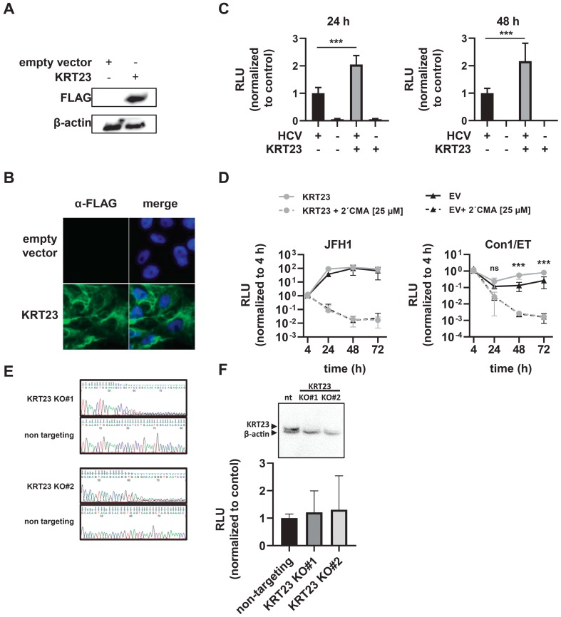
Expression of Keratin 23 facilitates HCV life cycle progression. (A) Western blot of Huh-7.5 cells stably expressing an empty vector (empty vector Huh-7.5) or 3xFLAG-KRT23 (KRT23 Huh-7.5). (B) Subcellular localization of KRT23 in KRT23 expressing Huh-7.5 cells. Cells were stained for 3xFLAG-tagged KRT23 (green), and nuclear DNA was stained with DAPI (blue). (C) Infection levels in empty vector and KRT23-expressing Huh-7.5 cells upon infection with HCV reporter virus JcR2a. Cells were infected with HCV JcR2a, and replication levels were determined by Renilla luciferase activity assay 24 h and 48 h post-infection. (D) HCV RNA replication efficiency of subgenomic replicons JFH1 NS3-3′and Con1/ET in empty vector and KRT23 Huh-7.5 cells. The firefly luciferase activity assay was performed to determine HCV RNA replication at the indicated time points. (C,D) Depicted are mean values ± standard deviation from three independent experiments (one-way ANOVA adjusted with Dunnett’s multiple comparison test; * p < 0.05; *** p < 0.001; ns: non-significant). (E) Editing of the target sequences in the designated KO cell lines. For validation, genomic DNA was sequenced to validate the loss of the consensus sequence in KO cell lines, compared with the non-targeting control. (F) Western blot of KRT23 KO Huh-7.5 cells and infection levels in non-targeting and KRT23 KO Huh-7.5 cells upon infection with HCV reporter virus JcR2a. Cells were infected with HCV JcR2a, and replication levels were determined by Renilla luciferase activity assay 48 h post-infection. (C,D,F) Depicted are mean values ± standard deviation from three independent experiments (one-way ANOVA adjusted with Dunnett’s multiple comparison test; * p < 0.05; *** p < 0.001; ns: non-significant).

