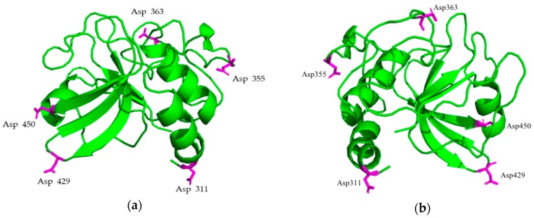
Figure 1.
Mutation sites of PlyC CHAP. (a) 3.3 Å resolution of the PlyC CHAP crystal structure. The magenta-colored amino acids represent the potential mutation sites. (b) 180° horizontal rotation of (a). The mutation sites are solvent exposed, not structured in α-helix or β-sheets, and form no interactions with other residues.

