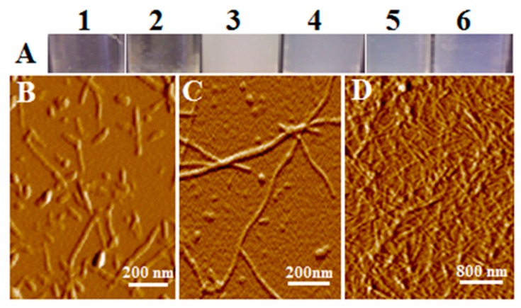
Figure 1.
Atomic force microscopy (AFM) images of lysozyme fiber solution and gel. (A) visual appearance of the fiber solution (1 and 2) and gel (3 to 6). Gels appear in two colors, white (vial 3) and light blue (vials 4–6). Fiber solution and gels were formed by incubation of lysozyme under different concentrations at 55 °C, and pH 2.5, adjusted with HCl (vial 1, 2, 5, 6) and pH 3.0 (vial 3 and 4). Vials (1) 10 mg/mL; (2) 20 mg/mL; (3) 40 mg/mL; (4) and (5) 70 mg/mL; (6) 100 mg/mL. (B–D) Representative AFM images are presented: (B) solution from vial 1; (C) solution of gel diluted in water; (D) gel. Colloidal spheres, beaded chains, and the unbranching, linear morphology of the early stage fibers shown in panels B and C suggest a linear aggregation pathway. Panel D reveals a dense network of fiber structure piled on top of each other.

