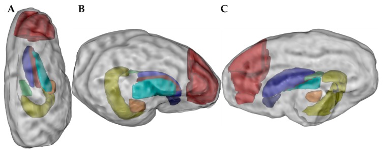
Figure 1.
Regions of interest (ROIs) after magnetic resonance imaging presented in top view (A), lateral view (B), and medial view (C), respectively. Measured ROIs: nucleus accumbens (dark blue), caudate nucleus (blue), lentiform nucleus (cyan), fornix (green), hippocampus (yellow), amygdala (orange), prefrontal cortex (red), and internal capsule (burgundy).

