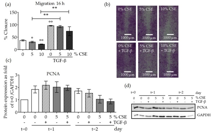
Figure 5.
CSE exposure decreased MSC migration and proliferation. In order to investigate the effect of CSE on SCP-1 cell migration, a scratch assay was performed. SCP-1 cells (N ≥ 3, n ≥ 3) were co-incubated with CSE (5–10%) and rhTGF-β1 (10 ng/mL). Wound closure was determined from microscopic pictures (equation: (100 − wound area at 16 h/wound area at 0 h) × 100) with ImageJ software. (a) SCP-1 cell migration after 16 h. (b) Representative migration pictures. Cells were visualized with sulforhodamine B (SRB) staining. (c) Normalized proliferating cell nuclear antigen (PCNA) protein expression in SCP-1 cells at day 0 (t = 0) and after 24 h (t = 1) or 48 h (t = 2) stimulation with rhTGF-β1 (10 ng/mL) and with or without CSE (5% v/v). (d) A representative PCNA Western blot. The results are expressed as mean ± SEM. Statistical significance was determined by the Kruskal–Wallis H test, followed by Dunn’s post-test. Significance was established as ** p < 0.01 compared to TGF-β1-treated cells and ° p < 0.05 or °° p < 0.01 compared to untreated cells.

