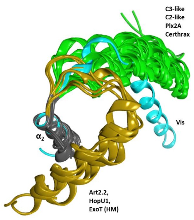
Figure 18.
Conformations of the α1 helix. Superposition of several toxins of the CT-group for the α2 helix (gray ribbons) showing three conformation clusters for the α1 helix. These clusters are the canonical conformation in green, the altered conformation of Vis in cyan, and the folded conformation of HopU1, Art2.2, and ExoT (HM) in ochre.

