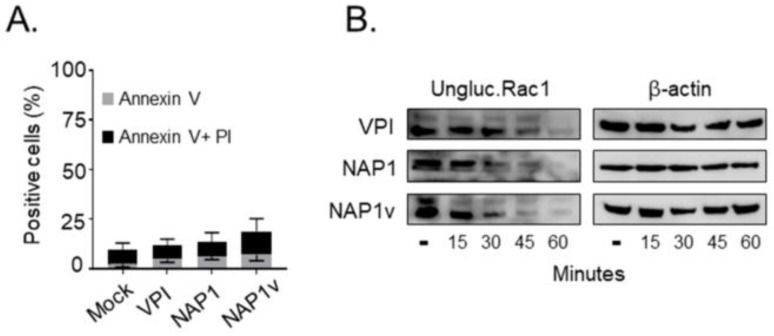
Figure 3.
Cytotoxicity of TcdBs in HeLa cells. (A) HeLa cells were treated with 100 pM of TcdBVPI (VPI), TcdBNAP1 (NAP1), and TcdBNAP1v (NAP1v) for 24 h. Cytotoxicity was analyzed by flow cytometry using propidium iodide (PI)/anexin V double staining. Control cells were left untreated (mock). Error bars represent means ± SD of three independent experiments; (B) Following the addition of 100 pM of TcdB, the cells were lysed at the indicated time points, and the dynamics of Rac1 glucosylation was monitored by immunoblot with a specific anti-Rac1 antibody that only recognizes the unmodified form of this protein (ungluc.Rac1). Untreated cells (-) were included as a positive control for unglucosylated Rac1, and immunodetection of beta-actin served as a loading control (β-actin). Shown are representative western blot images from three independent experiments.

