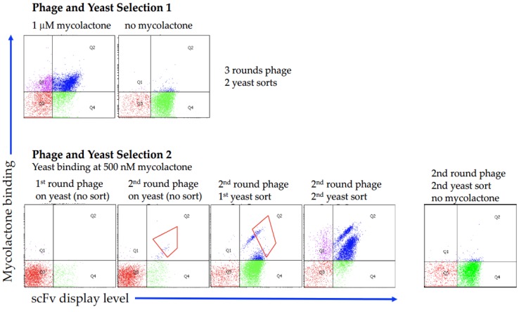
Figure 2.
Analysis of the flow cytometric output of scFv selections displayed on the surface of yeast. Selection 1 was carried out using plastic containers, while Selection 2 was carried out using glass containers. For Selection 2 the enrichment progress during the sorting steps is shown, including the gates used for the sorting of binding cells. The populations labeled as “no mycolactone” show the background binding for the fluorescently conjugated streptavidin.

