
Fig. 1.
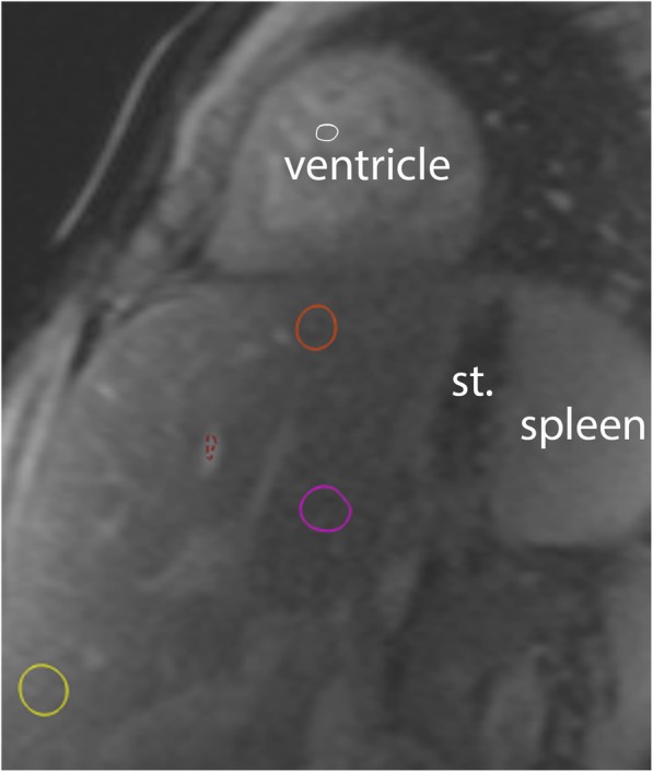
Hepatic Native T1. Native T1 source image in short-axis orientation demonstrating the heart and upper part of the abdomen. Regions of interest (ROIs) depict liver regions near diaphragm (#1, orange), central (#2, pink) and caudal (#3, yellow). The red ROI samples signal in the blood pool of a liver vein and the white ROI (#5) in the ventricular cavity. Stomach (st)