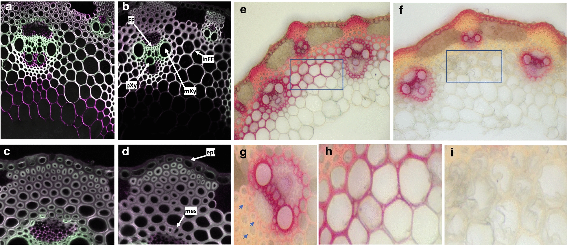
Fig. 5.

Two-photon fluorescence microscopy imaging and phloroglucinol–HCl staining of WT and lac5 lac8 lignified tissues. Cross sections of 30-day-old plants were imaged using two-photon fluorescence microscopy (green: 420–460-nm emission, purple: 495–540-nm emission a–d) or stained with phloroglucinol–HCl prior imaging under visible microscopy (e–i). WT: a, c, e, h; lac5 lac8: b, d, f, g, i. FF intrafascicular fibers, pXy protoxylem, mXy metaxylem, inFF interfascicular fibers, epi epidermis, mes mestome. Blue arrows show red staining of primary cell wall between interfascicular fiber cells in the double mutant
