Figure 1.
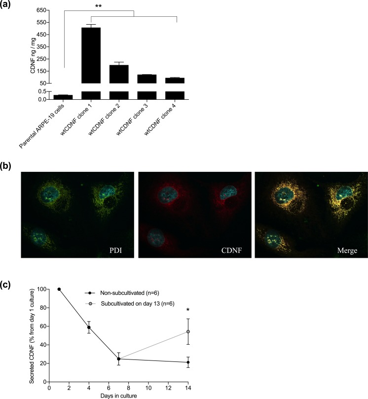
High levels of intracellular CDNF, which localize to the ER in the stable wtCDNF ARPE-19 cell clones. (a) Intracellular CDNF concentration in the clones ranged from 95.9 ± 2.9 to 508.3 ± 25.1 ng/mg (mean ± SEM, n=2–3/clone), whereas, in the parenteral ARPE-19 cell line it was 0.28 ± 0.01 ng/mg total protein (n=4). The increase in the clone intracellular CDNF concentration was up to 1800-fold compared with the parenteral cell line (p=0.001–0.007, one-way ANOVA + Dunnett’s test). (b) Immunofluorescent image of a wtCDNF ARPE-19 cell clone double stained to show the ER (PDI, green) and CDNF (red) and imaged by confocal microscopy. Merged image shows co-localization of CDNF and ER-marker (yellow). (c) CDNF secretion decreased down to 21.2 ± 5.7% from the initial secretion when the wtCDNF ARPE-19 clones were held without sub-cultivation for 14 days. Parallel to non-subcultivated, the same clones were subcultivated 1:2 on day 13 and CDNF secretion was analyzed at day 14. In the subcultivated cells CDNF secretion was 54.3 ± 13.8% from the initial secretion (p=0.036, paired samples t-test, n=6). ** p<0.01, * p<0.05.

